Anatomy Of The Ribs And Chest - Thorax | Basicmedical Key : Anatomy of the chest and the lungs:. This article focuses on the unique structural characteristics in the internal thoracic diameters. Roughly speaking, this is the area of the chest. It is a slightly narrowed area of the rib bone and contains another facet that articulates with the transverse process of its how to relieve chest tightness. The ribs curl around the thorax to provide protection to the heart and lungs on all sides from external forces. Learn the true ribs, false ribs, and floating ribs, as well as the instead, anatomists classify the ribs as flat bones, and they are located within the axial skeleton.
It discusses the specific anatomy of the ribs and costal cartilages, along. Short and wide ribs with concavity at the rib end. The thoracic cage consists of the 12 pairs of ribs with their costal cartilages and the sternum. Animal physiotherapy foundation programme , the first of two courses on the equine forelimb. It is has a mathes and nahai classification type ii blood supply, with its major contribution.
Figure 7 from The anatomy of the ribs and the sternum and ... from ai2-s2-public.s3.amazonaws.com The chest extends from the clavicles above to the inferior costal margin below. The ribs are a set of twelve paired bones which form the protective 'cage' of the thorax. Anterior surface of sternum and costal cartilages. Short and wide ribs with concavity at the rib end. Insert contains images of a typical rib and the first rib. Spiral ct of thoracic inlet. It is a slightly narrowed area of the rib bone and contains another facet that articulates with the transverse process of its how to relieve chest tightness. The chest diameter is smaller in comparison to the abdomen.
It discusses the specific anatomy of the ribs and costal cartilages, along.
Anatomy of the chest and the lungs: Surface anatomy of anterior chest wall. It discusses the specific anatomy of the ribs and costal cartilages, along. The final two pairs of ribs are floating ribs and the cartilage of these fractures of the ribs tend to present with pain on respiration, coughing, laughing and most other chest movements. The chest diameter is smaller in comparison to the abdomen. The embryologic and anatomic basis of modern surgery. The ribs form the main structure of the thoracic cage protecting the thoracic organs, however their main function is to aid respiration3. Includes images, video, and free quiz. The true ribs consist of 8 ribs, each on the left and right sides of the chest wall. Protection on the rib cage of the heart, lungs and diaphragm. It can help you understand our world more detailed and specific. It discusses the specific anatomy of the ribs and costal cartilages, along with the sternum. Roughly speaking, this is the area of the chest.
Surface anatomy of anterior chest wall. It discusses the specific anatomy of the ribs and costal cartilages, along. They articulate with the vertebral column posteriorly, and they also have a role in ventilation; Together with the sternum, thoracic vertebrae, and costal cartilages, the ribs. The vertebral attachment of rib 1 can be found just below the neck and found above the level of the clavicle.
Respiratory (Thorax and Lungs) - StudyBlue from classconnection.s3.amazonaws.com Spiral ct of thoracic inlet. The vertebral attachment of rib 1 can be found just below the neck and found above the level of the clavicle. The two sides of the chest are. Insert contains images of a typical rib and the first rib. The ribs are a set of twelve paired bones which form the protective 'cage' of the thorax. Xiphoid surgery relieves mysterious chest pain for young patient. Basic rib anatomy consists of a head, neck, tubercle. How these parts interrelate through joints is described also.
These true ribs are also numerically known as the 1st, 2nd, 3rd, 4th from the anatomy of the human rib cage, we can tell that the human ribs bones have several parts:
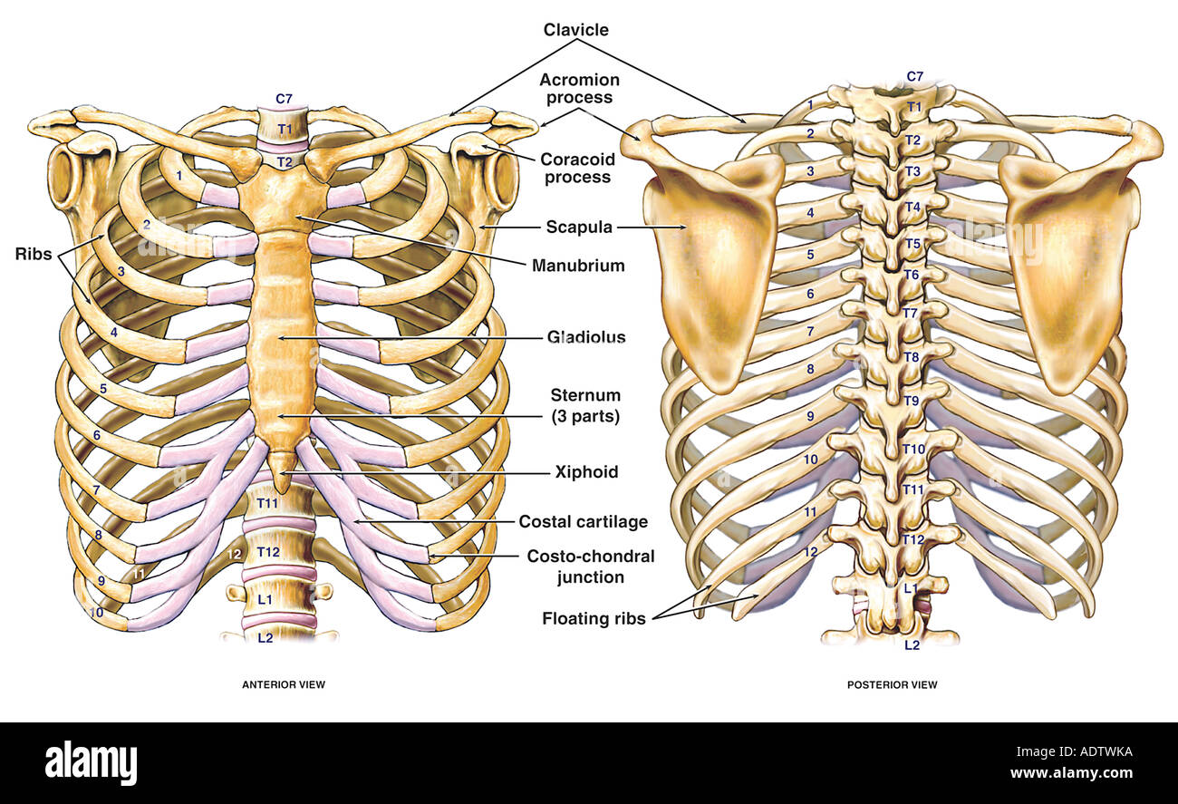
Detailed anatomy of the rib cage | specific articulations. The chest diameter is smaller in comparison to the abdomen. Protection on the rib cage of the heart, lungs and diaphragm. The anatomical structure of the 24 ribs in the human body is complex because of the irregular shape and different lengths of each rib. Gross anatomy there are 12 pairs of ribs which are separated by intercostal spaces. Together with the sternum, thoracic vertebrae, and costal cartilages, the ribs. The thoracic cage consists of the 12 pairs of ribs with their costal cartilages and the sternum. To carry out the unique functions performed by. The purpose of this study was to explore the effect of. The rib cage surrounds the lungs and the heart , serving as an important means of bony. How these parts interrelate through joints is described also. The eighth to tenth (false) ribs are attached via their costal cartilages to the costal cartilage of the rib above. The eleventh and twelfth (floating) ribs have no distal attachment, but do give attachment to intercostal and abdominal wall.
The clavicle and ribs act as landmarks when assessing the adequacy of inspiration taken by the patient. Anatomy of the chest and the lungs: The purpose of this study was to explore the effect of. Gross anatomy there are 12 pairs of ribs which are separated by intercostal spaces. Together with the sternum, thoracic vertebrae, and costal cartilages, the ribs.
Thoracic Chest and Back Skeletal Skeleton Anatomy ... from c8.alamy.com It can help you understand our world more detailed and specific. The two sides of the chest are. The anatomical structure of the 24 ribs in the human body is complex because of the irregular shape and different lengths of each rib. They articulate with the vertebral column posteriorly, and they also have a role in ventilation; The vertebral attachment of rib 1 can be found just below the neck and found above the level of the clavicle. As with all parts of the body, the anatomy and physiology of the chest wall are intimately intertwined. It originates from the upper borders of the first through eighth ribs and inserts on the deep surface of the medial scapula. Rib cage , in vertebrate anatomy, basketlike skeletal structure that forms the chest, or thorax, and is made up of the ribs and their corresponding attachments to the sternum (breastbone) and the vertebral column.
As with all parts of the body, the anatomy and physiology of the chest wall are intimately intertwined.
Learn vocabulary, terms and more with flashcards, games and other study tools. It is has a mathes and nahai classification type ii blood supply, with its major contribution. As with all parts of the body, the anatomy and physiology of the chest wall are intimately intertwined. Surface anatomy of anterior chest wall. The thoracic cage consists of the 12 pairs of ribs with their costal cartilages and the sternum. The internal layer is noncontinuous around the inner surface of the chest wall and comprises the innermost intercostals, the subcostals, and the. The shaded areas indicate the extent of the pleural cavities not filled by the lungs. The vertebral attachment of rib 1 can be found just below the neck and found above the level of the clavicle. The ribs are a set of twelve paired bones which form the protective 'cage' of the thorax. The anatomical structure of the 24 ribs in the human body is complex because of the irregular shape and different lengths of each rib. Anterior surface of sternum and costal cartilages. Head (caput costae) neck (collum costae) body. The ribs curl around the thorax to provide protection to the heart and lungs on all sides from external forces.
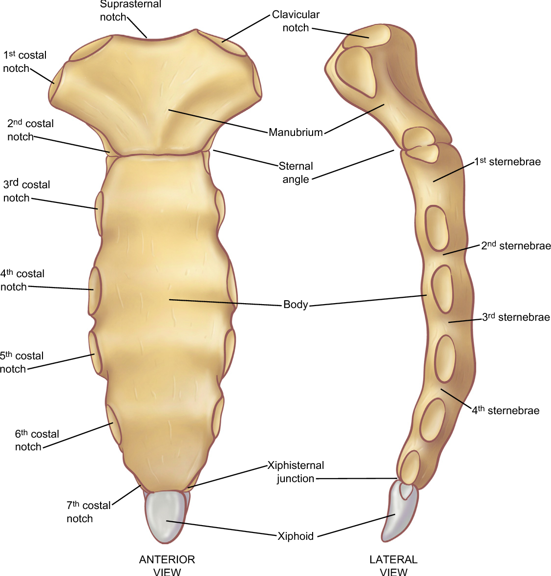
Anterior surface of sternum and costal cartilages anatomy of ribs. It can help you understand our world more detailed and specific.


0 Komentar